Mast Cells – Visual Perspectives

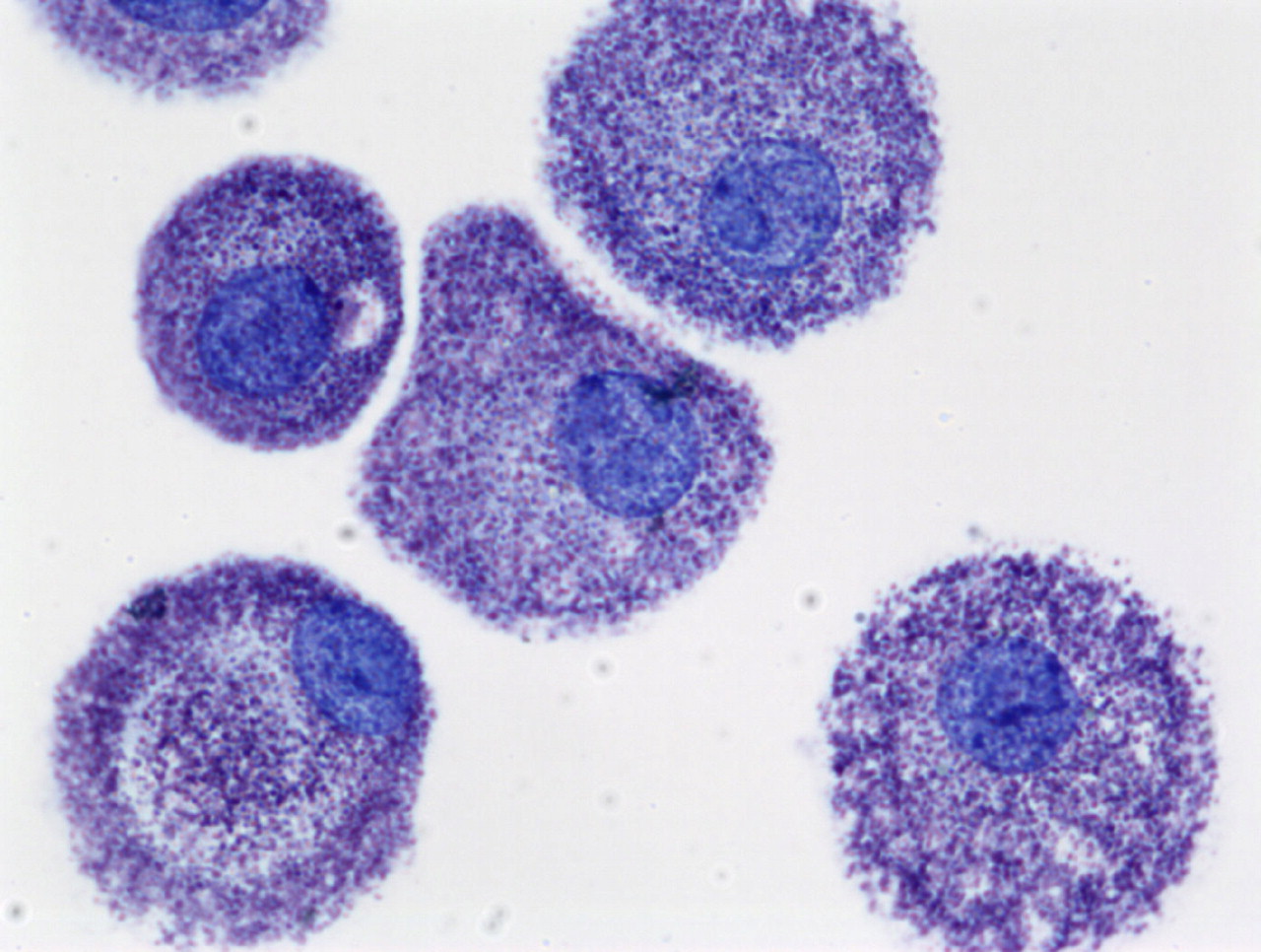
Mast cells are a part of the immune system and can be found throughout the body. Inside the mast cell are numerous secretory granules containing different chemicals that cause allergies and inflammation. These unique cells control many bodily functions and are important immune regulators and infection fighting cells. However, if we have too many of these covert immune cells and/or they misbehave, severe symptoms and serious disorders can result.

Mast Cells: Laboratory of Allergic Diseases, National Institute of Allergy and Infectious Diseases, National Institutes of Health (LAD/NIAID/NIH)

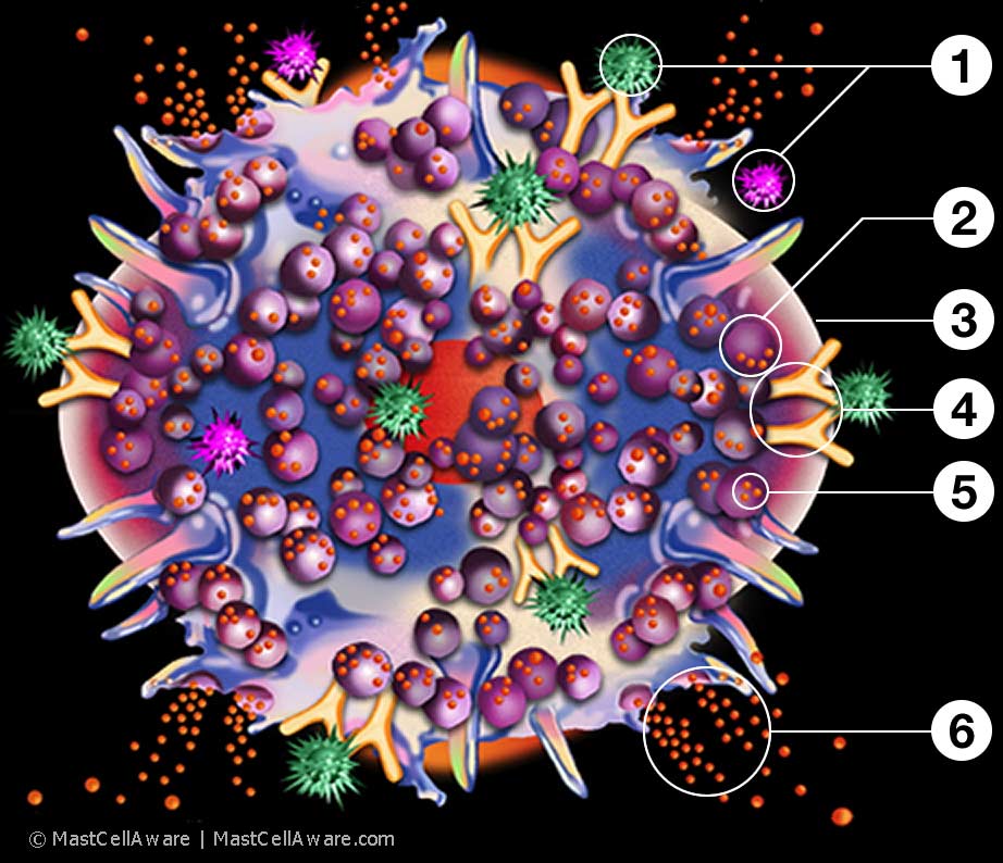
When allergens, drugs, toxins, etc. (green & pink spiked shapes) bind to the surface of the mast cell or cross-bridge the IgE antibodies (yellow Y-shaped pairs), the mast cell is triggered releasing histamine, tryptase and other mediators into the body (represented by the release of orange granules) leading to localized or systemic symptoms.

The illustration below is a graphic interpretation of the mast cell in activation and it's main components.

Mast Cells: Laboratory of Allergic Diseases, National Institute of Allergy and Infectious Diseases, National Institutes of Health (LAD/NIAID/NIH)

Mast Cells In Activation

Click the links to learn more about the parts of a mast cell. (Links open in new window)

In this illustration, the mast cell, similar in appearance to a white blood cell (white/blue areas), contains many storage granules (purple spheres) rich in histamine and tryptase (small orange granules).When allergens, drugs, toxins, etc. (green & pink spiked shapes) are binding & cross-bridging the antibodies (yellow Y-shaped pairs), a mast cell reaction is triggered, releasing histamine, tryptase and other mediators into the system (represented by the release of orange granules).

Thanks to Theoharis C. Theoharides, MS, PhD, MD, FAAAAI for his guidance with this visual interpretation of mast cells in activation.